Immediate Loading Dental Implants Protocol: 7 Key Steps by Dr. Ali Cem Ulubey
Quick answer: Immediate loading means fixed temporary teeth are attached to new implants on the day of surgery. At Ulubey Dental Clinic in Antalya, Dr. Ali Cem Ulubey and Dr. Surur Ulubey do this only when the implants are stable enough: an insertion torque of at least 35–45 Ncm and an ISQ of 68–70, which they measure. If stability is lower, for example in soft bone, the implants heal for about 3 months before loading. Temporary teeth are fitted within about 2–3 hours, followed by a soft-food diet during healing.
Executing an evidence-based immediate loading dental implants protocol allows patients undergoing full-arch or anterior tooth replacement to receive functional, aesthetic fixed temporary teeth on the very day of surgery. However, in contemporary implantology, same-day teeth must never be treated as a marketing gimmick or a universal guarantee.
Whether you are considering our patient-focused same-day dental implants guide or evaluating definitive restorative surgery like All-on-4 dental implants in Antalya and All-on-6 dental implants, immediate loading requires strict adherence to primary mechanical stability and biological healing kinetics. This clinical guide breaks down the 7 key steps of our immediate loading protocol at Ulubey Dental Clinic in Antalya, combining objective resonance frequency diagnostics (ISQ ≥ 68–70), BTI Endoret PRGF autologous plasma acceleration, and same-day Formlabs 3D-printed temporaries.
Quantitative benchmarks for an immediate loading dental implants protocol established in peer-reviewed implant literature (Albrektsson, Cochran, Morton) and clinical practice in Antalya:
- Step 1: Primary Mechanical Stability vs. Secondary Biological Osseointegration
- Step 2: The Critical Micromotion Threshold (< 150 µm Physics)
- Step 3: Objective Resonance Frequency Diagnostics (ISQ ≥ 68–70)
- Step 4: Accelerating Osteogenesis with BTI Endoret PRGF Plasma
- Step 5: Same-Day 3D-Printed Temporaries via Formlabs Workflow
- Step 6: Rigid Cross-Arch Splinting & Passive Occlusal Scheme
- Step 7: Managing the 3-to-5 Week Biological “Stability Dip”
- Clinical FAQs: Pain, Candidacy & Travel Guidelines
- Peer-Reviewed Implant Literature Citations
Step 1: Primary Mechanical Stability vs. Secondary Biological Osseointegration
To safely execute an immediate loading dental implants protocol, clinicians must differentiate between two concurrent stability phases described in classical bone biology:
- Primary Stability (Mechanical): Achieved on the day of surgery via mechanical friction between the titanium implant threads and the surrounding cortical and trabecular osteotomy walls. It is purely physical and begins decaying immediately as surgical micro-fractures undergo osteoclastic resorption.
- Secondary Stability (Biological): Developed over 4 to 12 weeks as newly differentiated osteoblasts lay down woven and lamellar bone directly onto the titanium oxide layer (Albrektsson et al., 1981).
Immediate loading is only biologically viable when initial primary mechanical stability is sufficiently robust to anchor the fixture while secondary biological bone remodeling takes over. Review our comparative analysis in our All-on-4 vs All-on-6 clinical guide.
Step 2: The Critical Micromotion Threshold (< 150 µm Physics)
Why do implants fail when loaded prematurely? The biological answer lies in cellular mechanotransduction at the microscopic bone-implant boundary:
Δx > 150 μm → Fibroblastic Encapsulation (Failure)
• Δx: Interfacial micromotion between titanium implant surface and alveolar bone under functional load.
• Physiological Tolerance: Micromotion below 100 µm stimulates bone remodeling.
• Pathological Threshold: Micromotion exceeding 150 µm ruptures immature capillary buds, depriving pluripotent mesenchymal cells of oxygen and causing fibrous scar encapsulation instead of bone (Szmukler-Moncler et al., 1998).
Our immediate loading dental implants protocol is engineered specifically to maintain interfacial micromotion well below this 100 µm threshold through high insertion torque, undersized osteotomy site preparation, and rigid cross-arch splinting.
Step 3: Objective Resonance Frequency Diagnostics (ISQ ≥ 68–70)
Subjective tactile feeling with a hand ratchet is inadequate for determining immediate loading suitability. At Ulubey Dental Clinic, Dr. Ali Cem Ulubey evaluates every fixture using Resonance Frequency Analysis (RFA) to calculate the precise Implant Stability Quotient (ISQ):
| ISQ Value Range | Clinical Stability Status | Loading Protocol Decision |
|---|---|---|
| ISQ < 60 | Low Primary Stability | Immediate Loading Contraindicated: Submerge fixture or place healing abutment; 3-month delayed healing. |
| ISQ 60 – 67 | Moderate Stability | Case-Specific Evaluation: Suitable for progressive loading only if rigidly splinted to multiple high-torque implants. |
| ISQ ≥ 68 – 70+ | High Mechanical Stability | Immediate Loading Approved: Proceed with immediate 3D-printed screw-retained provisional restoration. |
A magnetic resonance transducer is attached to the implant fixture or multi-unit abutment. The device emits magnetic pulses that induce micro-resonance, translating the response into an Implant Stability Quotient (ISQ) on a calibrated scale from 1 to 100 (Meredith, 1998). This provides a repeatable, scientifically validated baseline before a single tooth is loaded.
Step 4: Accelerating Osteogenesis with BTI Endoret PRGF Plasma
To reduce post-operative inflammation and accelerate biological bone deposition, we integrate BTI Endoret PRGF (Plasma Rich in Growth Factors) into our surgical protocol:
Immediately prior to surgery, a small sample of the patient’s venous blood is centrifuged in a specialized BTI Endoret unit. This separates leukocyte-free plasma rich in concentrated autologous growth factors, including:
- Platelet-Derived Growth Factor (PDGF): Stimulates osteoblast and fibroblast replication.
- Transforming Growth Factor-Beta (TGF-β): Promotes collagen matrix deposition and bone mineralization.
- Vascular Endothelial Growth Factor (VEGF): Accelerates angiogenesis, establishing new blood supply around the fixture (Anitua et al., 2007).
We coat the titanium implant surface with liquid PRGF prior to seating and use PRGF fibrin membranes to seal the surgical alveolus, reducing soft-tissue edema and accelerating peri-implant mucosal sealing.
Step 5: Same-Day 3D-Printed Temporaries via Formlabs Digital Workflow
Traditional chairside temporization involves messy acrylic dough (PMMA) that generates high exothermic heat (exceeding 60°C), risking thermal necrosis to the fresh osteotomy. In our digital protocol:
-
Digital Optical Impression
Immediately following implant placement, our Dentsply Sirona Primescan Connect intraoral scanner captures the exact soft-tissue emergence and multi-unit positions in minutes.
-
CAD Architecture in exocad
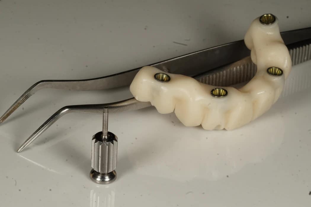
Dr. Surur Ulubey designs the monolithic provisional bridge, ensuring zero contact in eccentric excursions and creating gentle, convex mucosal contact areas that preserve the gingival architecture.
-
Formlabs Medical-Grade 3D Printing
The bridge is 3D-printed in-house using biocompatible restorative resin on a Formlabs stereolithography printer in approximately 90 minutes.
-
Passive In-House Delivery
Within 2 to 3 hours of surgery, the patient receives a fixed, aesthetic screw-retained smile. Zero messy putty impressions, zero heat generation, and zero delay.
Step 6: Rigid Cross-Arch Splinting & Passive Occlusal Scheme
In full-arch cases, immediate loading is made possible by cross-arch stabilization. By rigidly connecting 4 to 6 implants with a continuous framework, bite forces are distributed throughout the polygon of support, preventing any single fixture from enduring dangerous bending moments.
Our prosthetic design rules for immediate temporaries include:
- No Cantilevers: Provisional bridges terminate strictly at the most distal implant; posterior extension cantilevers are completely eliminated during the initial 12 weeks.
- Flat Occlusal Tables: Cusp angles are reduced to minimize lateral shear forces.
- Anterior Disclusion: Centric contacts are light, and canine guidance completely discludes posterior teeth in lateral excursions.
Step 7: Managing the 3-to-5 Week Biological “Stability Dip”
A critical biological phenomenon that patients must understand is the Stability Dip:
• Weeks 3–5 (The Critical Dip): Primary mechanical stability has dropped by ~30–40%, while newly formed woven bone has not yet fully mineralized. This is the highest risk window for implant failure.
• Weeks 6–12: Lamellar bone deposition peaks; secondary biological stability reaches maximum strength.
Because the implant enters this biological dip between weeks 3 and 5, patients must maintain a strict soft-food diet for 8 to 12 weeks. Once secondary osseointegration is confirmed, definitive restorations—such as our precision-milled monolithic zirconia frameworks—can be delivered with absolute peace of mind.
Clinical FAQs: Pain, Candidacy & Travel Guidelines
Peer-Reviewed Medical Literature Citations
- Albrektsson, T., et al. (1981). Osseointegrated titanium implants. Requirements for ensuring a long-lasting, direct bone-to-implant anchorage in man. Acta Orthopaedica Scandinavica, 52(2), 155–170.
- Szmukler-Moncler, S., et al. (1998). Timing of loading and effect of micromotion on bone-dental implant interface: a review of experimental literature. Journal of Biomedical Materials Research, 43(2), 192–203.
- Anitua, E., et al. (2007). The potential impact of the preparation rich in growth factors (PRGF) in different medical fields. Biomaterials, 28(31), 4551–4560.
- Meredith, N. (1998). Assessment of implant stability as a prognostic determinant. International Journal of Prosthodontics, 11(5), 491–501.
- Cochran, D. L., Morton, D., & Weber, H. P. (2004). Consensus statements and recommended clinical procedures regarding loading protocols for endosseous dental implants. International Journal of Oral & Maxillofacial Implants, 19(Suppl), 109–113.
- Morton, D., et al. (2018). Group 2 ITI Consensus Report: Prosthodontics and implant dentistry. Clinical Oral Implants Research, 29(Suppl 16), 215–223.
- Esposito, M., et al. (2013). Interventions for replacing missing teeth: different times for loading dental implants. Cochrane Database of Systematic Reviews, 2013(3), CD003878.
- Sennerby, L., & Meredith, N. (2008). Implant stability measurements using Resonance Frequency Analysis: biological and biomechanical aspects and clinical implications. Periodontology 2000, 47(1), 51–66.
- Misch, C. E., et al. (2004). Workshop guidelines on immediate loading in implant dentistry. Journal of Oral Implantology, 30(5), 283–288.
Last updated: · Editor contact: [email protected] · ulubeydental.com/contact-us