Dental X-Rays and CBCT: Why, How Much Radiation, What We Take
Dental X-rays show the bone, the roots and the inside of the teeth, which an examination cannot see. The doses are low: a panoramic X-ray of both jaws is about 0.01 mSv, roughly 1.5 days of natural background radiation in the UK[1]. A 3D CBCT scan gives more information and a higher dose, on average 0.08–0.21 mSv for adults depending on the field of view[2], so it is used only when a decision needs it[3]. At Ulubey Dental Clinic in Antalya every patient has a panoramic X-ray at the examination, with no separate fee. We do not take dental X-rays during pregnancy.
- 1. Why dentists take X-rays
- 2. Types of dental X-rays
- 3. How much radiation? Doses in perspective
- 4. When an X-ray is needed, and when not
- 5. CBCT: when a 3D scan helps
- 6. Pregnancy, breastfeeding and children
- 7. X-rays at our clinic
- 8. Sending your X-rays and getting copies
- 9. Frequently asked questions
- 10. Sources
Why dentists take X-rays
An X-ray is a test that takes pictures of the inside of the body using a small amount of radiation[4]. In dentistry it shows what a look into the mouth cannot: the bone around the roots, the area around the root tip, the inside of the teeth, and teeth that have not come through, such as wisdom teeth.
According to the 2026 recommendations of the American Dental Association and the American Academy of Oral and Maxillofacial Radiology, imaging decisions should be based on the patient’s medical and dental history, the clinical examination, the assessment of disease risk and specific clinical conditions. Used appropriately, X-rays contribute to treatment decisions and to good care[3].
Types of dental X-rays
| Type | What it shows | Typical effective dose |
|---|---|---|
| Small intraoral X-ray (bitewing or periapical) | One to a few teeth in detail; a periapical X-ray shows one to three teeth from the crown to the root tip, with the bone around them | Single bitewing: under 0.01 mSv[1] |
| Panoramic X-ray | All the teeth, both jaws, the jaw joints and the sinuses in one image | 0.01 mSv[1] |
| CBCT (cone-beam CT) | A 3D image of the jaw: bone height and width, the nerve canal and the sinus | Adults, mean: 84 µSv small field, 177 µSv medium, 212 µSv large (range 5–1,073 µSv)[2] |
| Medical CT of the head (for comparison) | Not a dental routine examination | About 2 mSv[1] |
1 mSv = 1,000 µSv. Doses vary with the machine and settings; the CBCT values are means from published data and nine scanners.
How much radiation? Doses in perspective
We are all exposed to natural background radiation. In the UK the average is 2.1 mSv per year, with regional averages from 1.5 to 7.5 mSv[1]. The UK Health Security Agency puts medical X-rays in that context:
| Examination | Typical dose | Equivalent natural background | Lifetime additional risk of fatal cancer |
|---|---|---|---|
| Teeth: single bitewing | < 0.01 mSv[1] | < 1.5 days | 1 in a few million |
| Teeth: panoramic | 0.01 mSv[1] | 1.5 days | 1 in 2 million |
| Chest X-ray (single film) | 0.02 mSv[1] | 3 days | 1 in a million |
| Dental CBCT, adult mean (small to large field) | 0.084–0.212 mSv[2] | About 15–37 days (calculated) | Not listed by UKHSA |
| CT head | 2 mSv[1] | 1 year | 1 in 10,000 |
CBCT days are our calculation from the Ludlow means and the UK average of 2.1 mSv per year; they are an orientation, not a measurement. UKHSA notes that for children the risk figures should be multiplied by about 2[1].
The NHS notes that having many X-rays carries a very small risk that the radiation could cause cancer, which is why the clinician assesses the risks before offering an X-ray[4]. Each X-ray should answer a clinical question.
When an X-ray is needed, and when not
There is no fixed schedule that suits everyone. The ADA and AAOMR recommend[3]:
- a thorough review of the patient’s history and the clinical findings before any X-ray;
- reviewing previously taken images first;
- using every imaging method, especially CBCT, judiciously, to keep the cumulative dose low.
In practice that means: bring or send your recent X-rays, tell us about scans taken elsewhere, and expect the dentist to explain why an X-ray is needed and what it will decide.
CBCT: when a 3D scan helps
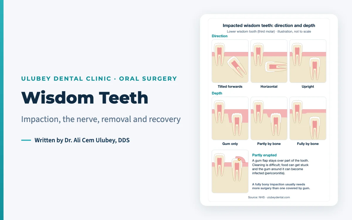
A CBCT scan shows bone height and width, the nerve canal and the sinus in three dimensions, which a flat panoramic image cannot. Typical reasons are planning implants, a bone graft or a sinus lift, and checking how close the roots of a lower wisdom tooth lie to the nerve.
CBCT is a planning tool, not a guarantee. A 2022 systematic review of seven randomised trials found moderate-quality evidence that adding CBCT does not routinely reduce nerve injury after high-risk lower wisdom tooth surgery[5]. Its value is in showing the anatomy so that the risk can be explained and the approach planned; see wisdom teeth. Because its dose is higher than a panoramic X-ray[1][2], it is taken when the answer will change the plan[3].
For implants, see dental implants in Antalya and dental bone graft.
Pregnancy, breastfeeding and children
At our clinic we do not take dental X-rays during pregnancy. During pregnancy we only do urgent work, in the second trimester and approved by your physician; definitive treatment is postponed until after the birth.
Please tell us if you are or might be pregnant, and if you are breastfeeding; the NHS asks patients to tell the healthcare professional about both before an X-ray[4]. More in our guide to oral health and medical conditions.
Children. Children are more sensitive to radiation: UKHSA suggests multiplying the lifetime risk figures by about 2 for children[1], and mean CBCT doses for children were 103 µSv (small field) and 175 µSv (large or medium field)[2]. The same rule applies as for adults, only more strictly: an X-ray only when it answers a question that matters for treatment[3]. Care for children at our clinic is described on our children’s dentist page.
X-rays at our clinic
Dr. Ali Cem Ulubey and Dr. Surur Ulubey examine every patient and read the images themselves; there are no rotating dentists.
| Image | When we take it |
|---|---|
| Panoramic X-ray (Vatech) | For every patient, at the examination, as the basis for diagnosis; included in the examination with no separate fee |
| Periapical X-ray | In the clinic, when a tooth needs a closer look, for example the root tip or a root canal |
| CBCT (3D) | When a case needs a 3D view of the bone or the nerve, for example implant planning or a lower wisdom tooth close to the nerve; evaluated by our dentists |
| During pregnancy | No dental X-rays |
The consultation has no fee. Any treatment that follows is itemised in your written quote, valid for 30 days.
The panoramic X-ray is also one of the documents for the yearly guarantee check-up: if you have the check-up with a dentist at home, you send us a panoramic X-ray (plus a periapical X-ray for implants, if taken), 5 standard intra-oral photos and the dentist’s note, and we review them within 7 days. See our written dental guarantee.
Sending your X-rays and getting copies
- Before you travel: send recent X-rays or clear photos of them on WhatsApp. The remote assessment and written quote have no fee; a written quote usually follows within 24 hours after the photos or X-rays arrive.
- Previous scans: tell us about any CBCT or X-rays taken elsewhere, so they can be reviewed first[3].
- Copies for your dentist: on request we share your records, such as the panoramic and periapical X-rays, with your dentist at home; see our treatment record handover checklist.
Frequently asked questions
Are dental X-rays safe?
Dental X-rays use very low doses. UKHSA gives 0.01 mSv for a panoramic X-ray, about 1.5 days of natural background radiation in the UK, and under 0.01 mSv for a single bitewing. Because any radiation carries a very small risk, an X-ray should only be taken when it answers a clinical question, and previous images should be reviewed first.
How much radiation is a dental CBCT scan?
More than a flat X-ray, and it varies widely by machine and field of view. A meta-analysis found mean adult doses of 84 µSv for small, 177 µSv for medium and 212 µSv for large fields, with a range from 5 to 1,073 µSv. That is why CBCT is used judiciously, when the 3D answer changes the treatment plan.
Do I need a CBCT scan for dental implants?
Often, but not always. A CBCT shows bone height and width, the nerve canal and the sinus in 3D, which matters for planning implants, a bone graft or a sinus lift. At our clinic every patient has a panoramic X-ray, and we use a CBCT scan when the case needs a 3D view of the bone; our dentists evaluate it.
Can I have a dental X-ray while pregnant?
At our clinic we do not take dental X-rays during pregnancy. We only do urgent work in the second trimester, approved by your physician, and postpone definitive treatment until after the birth. Please tell us if you are or might be pregnant, or if you are breastfeeding, before any examination.
Can I send my own X-rays before I travel?
Yes. Send recent X-rays or clear photos of them on WhatsApp, together with photos of your teeth. The remote assessment and written quote have no fee; the quote usually follows within 24 hours after the X-rays arrive. Reviewing existing images first is also what radiology guidance recommends.
How often should I have dental X-rays?
There is no fixed interval for everyone. Current ADA and AAOMR recommendations base the decision on your history, the examination and your risk of disease. Someone with frequent new decay or gum disease may need X-rays more often than someone with a stable, healthy mouth.
Sources
- UK Health Security Agency. Patient dose information: guidance. Medical radiation: patient doses (updated 28 September 2026; accessed 26 October 2026). www.gov.uk/government/publications/medical-radiation-patient-doses/patient-dose-information-guidance
- Ludlow JB, Timothy R, Walker C, et al. Effective dose of dental CBCT: a meta analysis of published data and additional data for nine CBCT units. Dentomaxillofac Radiol. 2015;44(1):20140197. PMID 25224586. pubmed.ncbi.nlm.nih.gov/25224586
- Benavides E, Krecioch JR, Allareddy T, Buchanan A, et al. American Dental Association and American Academy of Oral and Maxillofacial Radiology patient selection for dental radiography and cone-beam computed tomography. J Am Dent Assoc. 2026;157(1):20-35.e5. PMID 41500761. pubmed.ncbi.nlm.nih.gov/41500761
- NHS. X-ray (page last reviewed 4 February 2026; accessed 26 October 2026). www.nhs.uk/tests-and-treatments/x-ray
- Robbins J, Smalley KR, Ray P, Ali K. Does the addition of cone-beam CT to panoral imaging reduce inferior dental nerve injuries resulting from third molar surgery? A systematic review. BMC Oral Health. 2022;22(1):466. PMID 36329417. pubmed.ncbi.nlm.nih.gov/36329417
This article is general patient information and does not replace a clinical examination or X-rays.
Editor contact: [email protected] · ulubeydental.com/contact-us